| Image Records | |||||||||
| Image | Image ID | Figure title | Reference/ License | Sample ID/ Sample type/ Stage | Detection/ Microscope | Experiment/ Method | Gene | Keys | |
|---|---|---|---|---|---|---|---|---|---|
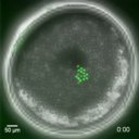 |
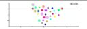
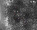
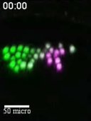
HO001B0002 | Cell behaviors during stripe formation in the spider Parasteatoda embryo | Hemmi et al.(2018) CC BY 4.0 |
NLS-tdEosFP_cellclone1 1_1 Ptep/embryo st_5_6_7_8 |
BF/NLS-tdEosFP/hh grayscale/green_red/green coolSNAP/confocal |
wild_type/cell_labeling/NLS-tdEosFP live/FISH |
aug3.g4322|hedgehog|hh|Pt-hh | time-lapse/extraembryonic | |
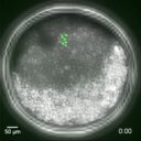 |
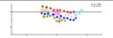
HO001B0003 | Cell behaviors during stripe formation in the spider Parasteatoda embryo | Hemmi et al.(2018) CC BY 4.0 |
NLS-tdEosFP_cellclone2 1_1 Ptep/embryo st_5_6_7_8 |
BF/NLS-tdEosFP/hh grayscale/green_red/green coolSNAP/confocal |
wild_type/cell_labeling/NLS-tdEosFP live/FISH |
aug3.g4322|hedgehog|hh|Pt-hh | time-lapse/opisthosoma/oscillation | |
 |
HO001B0004 | Cell behaviors during stripe formation in the spider Parasteatoda embryo | Hemmi et al.(2018) CC BY 4.0 |
NLS-tdTomato_cellclone 1_1 Ptep/embryo st_5_6_7 |
BF/NLS-tdEosFP grayscale/red coolSNAP |
wild_type/cell_labeling/NLS-tdTomato live |
time-lapse/opisthosoma/oscillation | ||
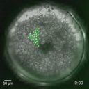 |
HO001B0005 | Cell behaviors during stripe formation in the spider Parasteatoda embryo | Hemmi et al.(2018) CC BY 4.0 |
H1-tdEosFP_cellclone 1_1 Ptep/embryo st_5_6_7 |
BF/H1-tdEosFP/noto1 grayscale/green_red/green coolSNAP/confocal |
wild_type/cell_labeling/H1-tdEosFP live/FISH |
aug3.g15243|noto1|not|Pt-noto1 | time-lapse/thorax/opisthosoma | |
 |
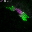
HO001B0006 | Cell behaviors during stripe formation in the spider Parasteatoda embryo | Hemmi et al.(2018) CC BY 4.0 |
H1-tdEosFP_cellclone 1_2 Ptep/embryo st_5_6_7 |
BF/H1-tdEosFP/noto1 many colors coolSNAP/confocal |
wild_type/cell_labeling/H1-tdEosFP live/FISH |
aug3.g15243|noto1|not|Pt-noto1 | tracking/animation/thorax/subdivision | |
 |
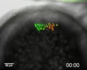
HO001B0007 | Cell behaviors during stripe formation in the spider Parasteatoda embryo | Hemmi et al.(2018) CC BY 4.0 |
H1-tdEosFP_cellclone 1_3 Ptep/embryo st_5_6_7 |
BF/H1-tdEosFP/noto1 many colors coolSNAP/confocal |
wild_type/cell_labeling/H1-tdEosFP live/FISH |
aug3.g15243|noto1|not|Pt-noto1 | tracking/animation/thorax/convergent_extension | |
 |
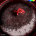
HO001C0003 | Cell behaviors during stripe formation in the spider Parasteatoda embryo | Hemmi et al.(2018) CC BY 4.0 |
microinjection_fate_map 1_1 Ptep/embryo st_5_6_7_8 |
BF/NLS-tdEosFP/hh grayscale/green_red/green confocal |
wild_type/cell_labeling/NLS-tdEosFP FISH |
aug3.g4322|hedgehog|hh|Pt-hh | cell_labeling | |
 |
HO001C0004 | Cell behaviors during stripe formation in the spider Parasteatoda embryo | Hemmi et al.(2018) CC BY 4.0 |
NLS-tdEosFP_cellclones_1 1_1 Ptep/embryo st_5_6_7_8 |
BF/NLS-tdEosFP/hh grayscale/green_red/green coolSNAP/confocal |
wild_type/cell_labeling/NLS-tdEosFP live/FISH |
aug3.g4322|hedgehog|hh|Pt-hh | cell_labeling | |
 |
HO001C0005 | Cell behaviors during stripe formation in the spider Parasteatoda embryo | Hemmi et al.(2018) CC BY 4.0 |
NLS-tdTomato_cellclone 1_1 Ptep/embryo st_5_6_7 |
BF/NLS-tdTomato grayscale/red coolSNAP |
wild_type/cell_labeling/NLS-tdTomato live |
cell_labeling/tracking | ||
 |
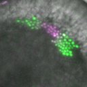
HO001C0006 | Cell behaviors during stripe formation in the spider Parasteatoda embryo | Hemmi et al.(2018) CC BY 4.0 |
H1-tdEosFP_cellclone 1_1 Ptep/embryo st_5_6_7 |
BF/H1-tdEosFP/noto1 grayscale/green/purple coolSNAP/confocal |
wild_type/cell_labeling/H1-tdEosFP live/FISH |
aug3.g15243|noto1|not|Pt-noto1 | cell_labeling/tracking | |
 |
HO001C0011 | Cell behaviors during stripe formation in the spider Parasteatoda embryo | Hemmi et al.(2018) CC BY 4.0 |
NLS-tdEosFP_cellclones_2 1_1 Ptep/embryo st_5_6_7_8 |
BF/NLS-tdEosFP/hh grayscale/green_red/green coolSNAP/confocal |
wild_type/cell_labeling/NLS-tdEosFP |
aug3.g4322|hedgehog|hh|Pt-hh | cell_labeling | |
 |
HO001C0012 | Cell behaviors during stripe formation in the spider Parasteatoda embryo | Hemmi et al.(2018) CC BY 4.0 |
NLS-tdEosFP_cellclones_3 1_1 Ptep/embryo st_5_6_7_8 |
BF/NLS-tdEosFP/hh grayscale/green_red/green coolSNAP/confocal |
wild_type/cell_labeling/NLS-tdEosFP |
aug3.g4322|hedgehog|hh|Pt-hh | cell_labeling | |
 |
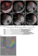
HO001D0001 | Traveling of a hh wave in the presumptive head region of the spider Parasteatoda embryo | Hiroki Oda (unpublished) |
H1tdEosFP_Head 1_1 Ptep/embryo st_5_6_7 |
BF/H1tdEosFP grayscale/green coolSNAP |
wild_type/cell_labeling/H1tdEosFP live |
aug3.g4322|hedgehog|hh|Pt-hh | time-lapse/cell_labeling/tracking/head/traveling | |
 |
HO001D0002 | Traveling of a hh wave in the presumptive head region of the spider Parasteatoda embryo | Hiroki Oda (unpublished) |
H1tdEosFP_Head 1_2 Ptep/embryo st_5_6_7 |
BF/H1tdEosFP/hh grayscale/green/red coolSNAP/confocal |
wild_type/cell_labeling/H1tdEosFP/tracking live |
aug3.g4322|hedgehog|hh|Pt-hh | time-lapse/cell_labeling/tracking/head/traveling | |
 |
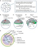
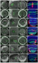
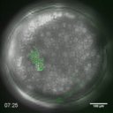
HO002A0013 | Cell internalization from the rim of the germ disc | Kanayama et al.(2011) CC BY-NC-SA 3.0 |
NLS-tdEosFP_GDrim 1_1 Ptep/embryo st_5_6_7 |
DIC/NLS-tdEosFP gray/green/red confocal |
wild_type/cell_labeling/NLS-tdEosFP/photo_conversion live |
time-lapse/mesoderm | ||
 |
HO002A0014 | Cell internalization from the rim of the germ disc | Kanayama et al.(2011) CC BY-NC-SA 3.0 |
NLS-tdEosFP_GDrim 1_2 Ptep/embryo st_5_6_7 |
NLS-tdEosFP green/purple confocal |
wild_type/cell_labeling/NLS-tdEosFP/photo_conversion live |
time-lapse/mesoderm | ||
 |
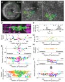
HO002A0015 | Convergent extension of the presumptive head ectoderm | Kanayama et al.(2011) CC BY-NC-SA 3.0 |
NLS-tdEosFP_Head 1_1 Ptep/embryo st_6_7 |
DIC/NLS-tdEosFP gray/green/red confocal |
wild_type/cell_labeling/NLS-tdEosFP/photo_conversion live/WISH |
time-lapse/head_ectoderm/convergent_extension/splitting | ||
 |
HO002A0016 | Convergent extension of the presumptive head ectoderm | Kanayama et al.(2011) CC BY-NC-SA 3.0 |
NLS-tdEosFP_Head 1_2 Ptep/embryo st_6_7 |
NLS-tdEosFP green/purple confocal |
wild_type/cell_labeling/NLS-tdEosFP/photo_conversion live/WISH |
time-lapse/head_ectoderm/convergent_extension/splitting | ||